
Labeled Pictures Of the Heart Lovely Simple Human Heart Diagram for Kids Human heart diagram
This diagram showcases the heart through the orientation of the frontal plane. The figure is labeled to showcase the heart's basic anatomical structures, including; right and left atria and ventricles, and the aorta.. This simplified schematic provides a visualization that may promote a better understanding. The diagram also does an.

Heart Diagram Sketch at Explore collection of Heart Diagram Sketch
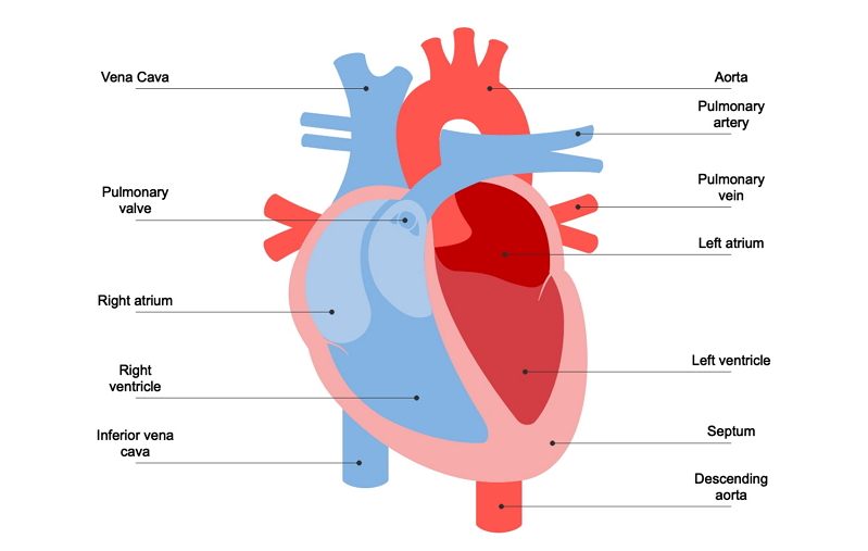
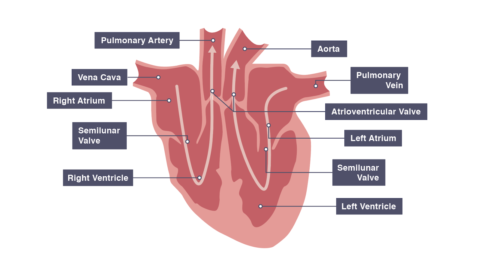
Figure 40.9. 1: Human Heart: (a) The heart is primarily made of a thick muscle layer, called the myocardium, surrounded by membranes. One-way valves separate the four chambers. (b) Blood vessels of the coronary system, including the coronary arteries and veins, keep the heart muscles oxygenated.

How to Draw the Internal Structure of the Heart (with Pictures)
Heart Your heart is the main organ of your cardiovascular system, a network of blood vessels that pumps blood throughout your body. It also works with other body systems to control your heart rate and blood pressure. Your family history, personal health history and lifestyle all affect how well your heart works.

humanheartdiagram Tim's Printables
The heart is a muscular organ that pumps blood around the body by circulating it through the circulatory/vascular system. It is found in the middle mediastinum, wrapped in a two-layered serous sac called the pericardium.

heart anatomy labeling
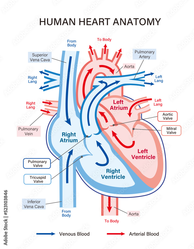
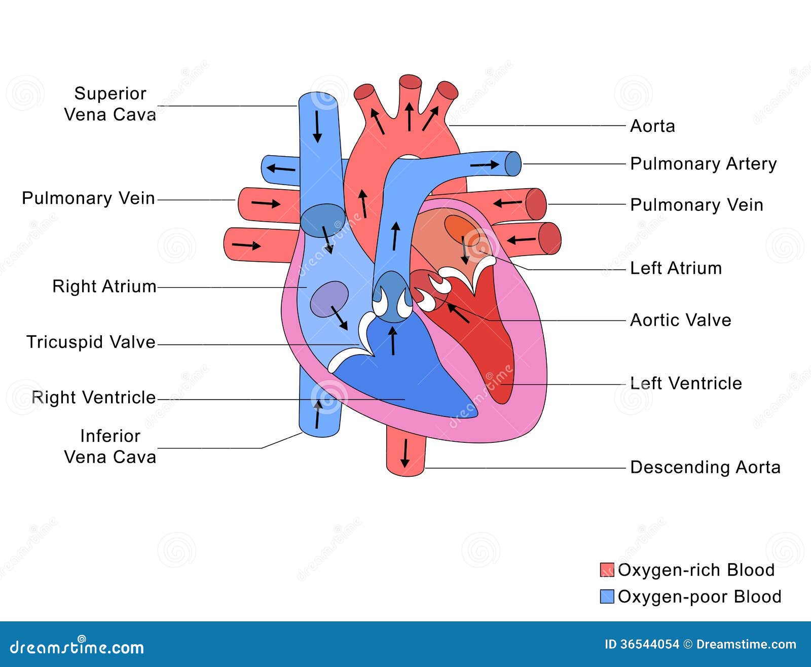
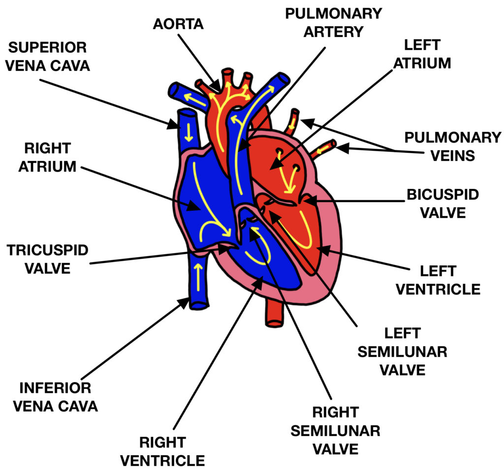
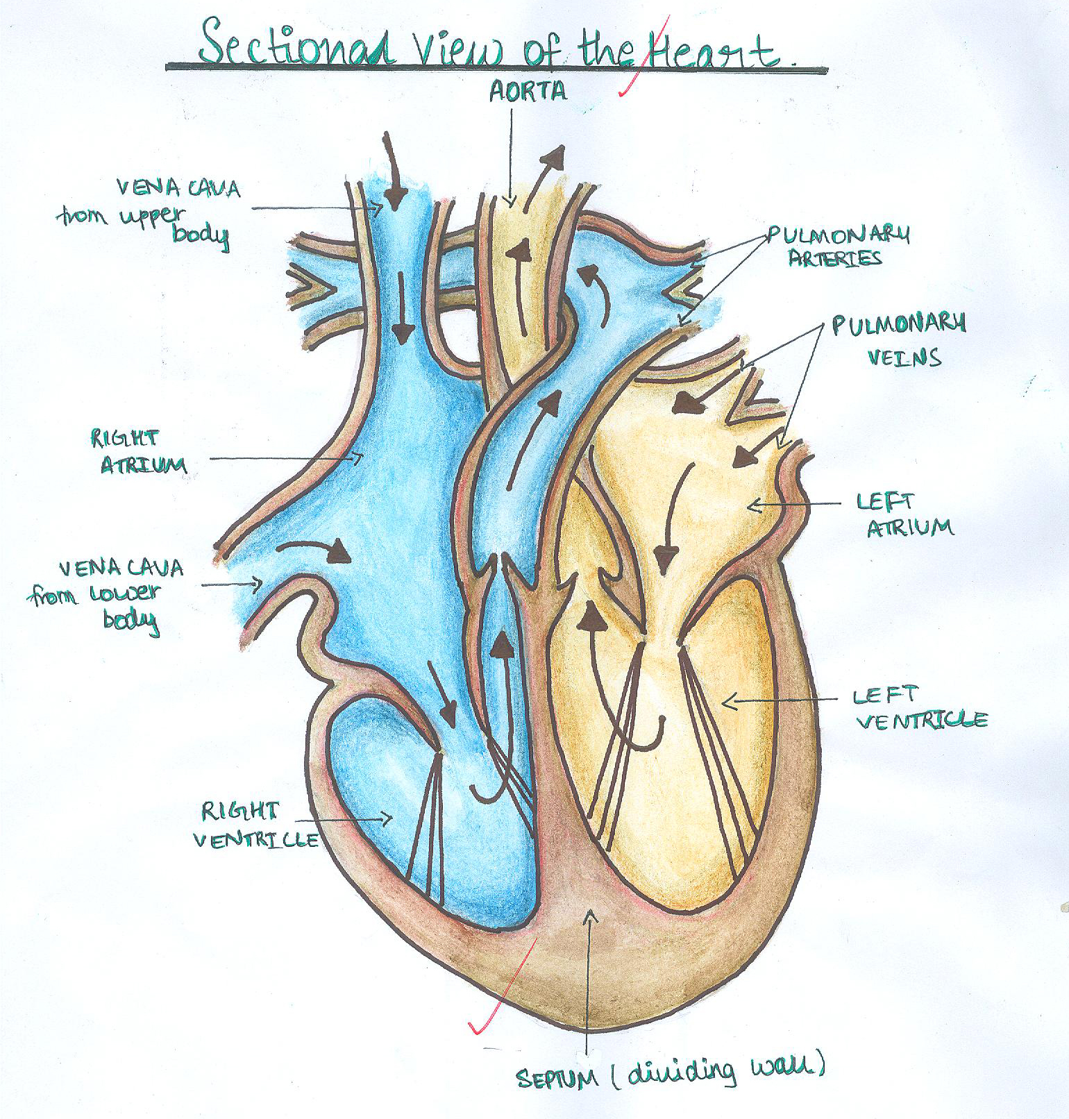
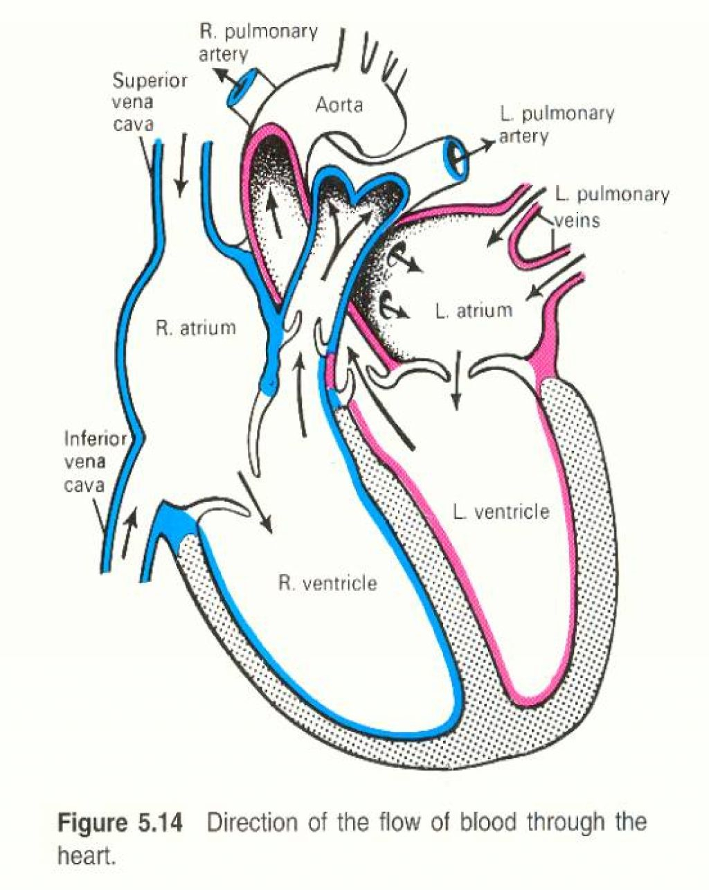
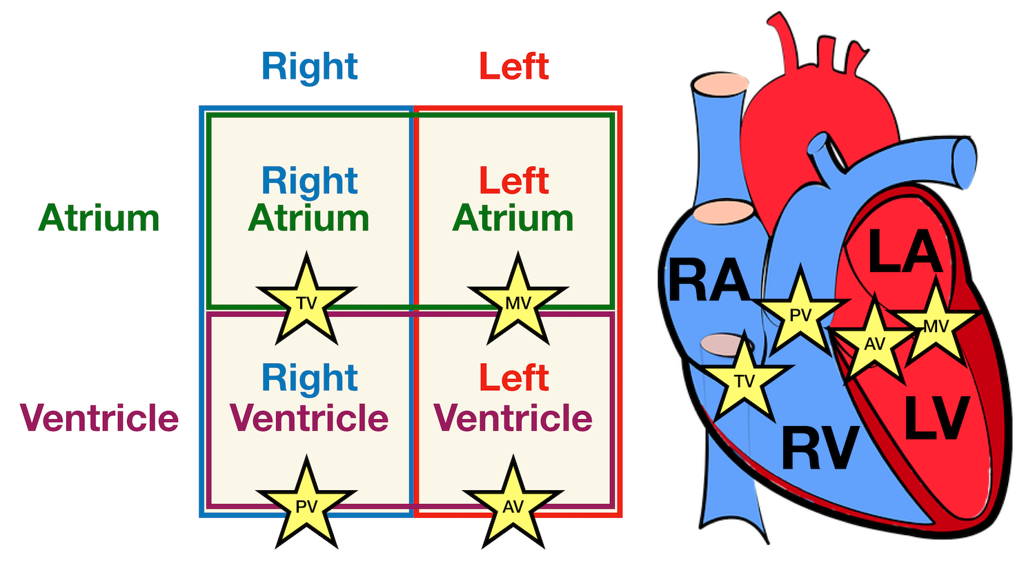
The human heart is primarily comprised of four chambers. The two upper chambers are called the atria, the remaining two lower chambers are the ventricles. The right and left sides of the heart are separated by a muscle called the "septum.". Both sides work together to efficiently circulate the blood.

Heart Anatomy Labeled Diagram, Structures, Blood Flow, Function of Cardiac System — EZmed
Diagram of Heart The human heart is the most crucial organ of the human body. It pumps blood from the heart to different parts of the body and back to the heart. The most common heart attack symptoms or warning signs are chest pain, breathlessness, nausea, sweating etc.
Human heart anatomy illustration explaining blood flow. A simple diagram, great for educational
The heart is an amazing organ. It starts beating about 22 days after conception and continuously pumps oxygenated red blood cells and nutrient-rich blood and other compounds like platelets throughout your body to sustain the life of your organs. Its pumping power also pushes blood through organs like the lungs to remove waste products like CO2.
human heart labelled diagram
This simple heart diagram with labels activity will help your pupils begin to understand the heart, what it does and the different parts that comprise it. The resource comes with two different diagrams of the heart; one with labels attached, and one blank diagram with the labels at the bottom for students to complete themselves. Ideal as an introductory lesson on the heart and how it.
IGCSE Biology 2017 2.65 Describe the Structure of the Heart and How it Functions
The structure of the heart If you clench your hand into a fist, this is approximately the same size as your heart. It is located in the middle of the chest and slightly towards the left. The.
Simplified Structure Of Heart Stock Images Image 36544054
The Heart is a muscular organ in the thoracic cavity that pumps blood through vessels in the body. It consists of four chambers: two atria and two ventricles. The heart is split into a left and a right side, with each side having one atrium and one ventricle. The interventricular septum divides the heart into the left and right ventricles, and.
Structure of the Heart The Science and Maths Zone
Higher; Structure and function of the heart The cardiac cycle. In this Higher Human Biology revision guide, you will learn in detail that cardiac output is a measure of the rate of blood flow.
Human Heart Drawing Simple at Explore collection of Human Heart Drawing Simple
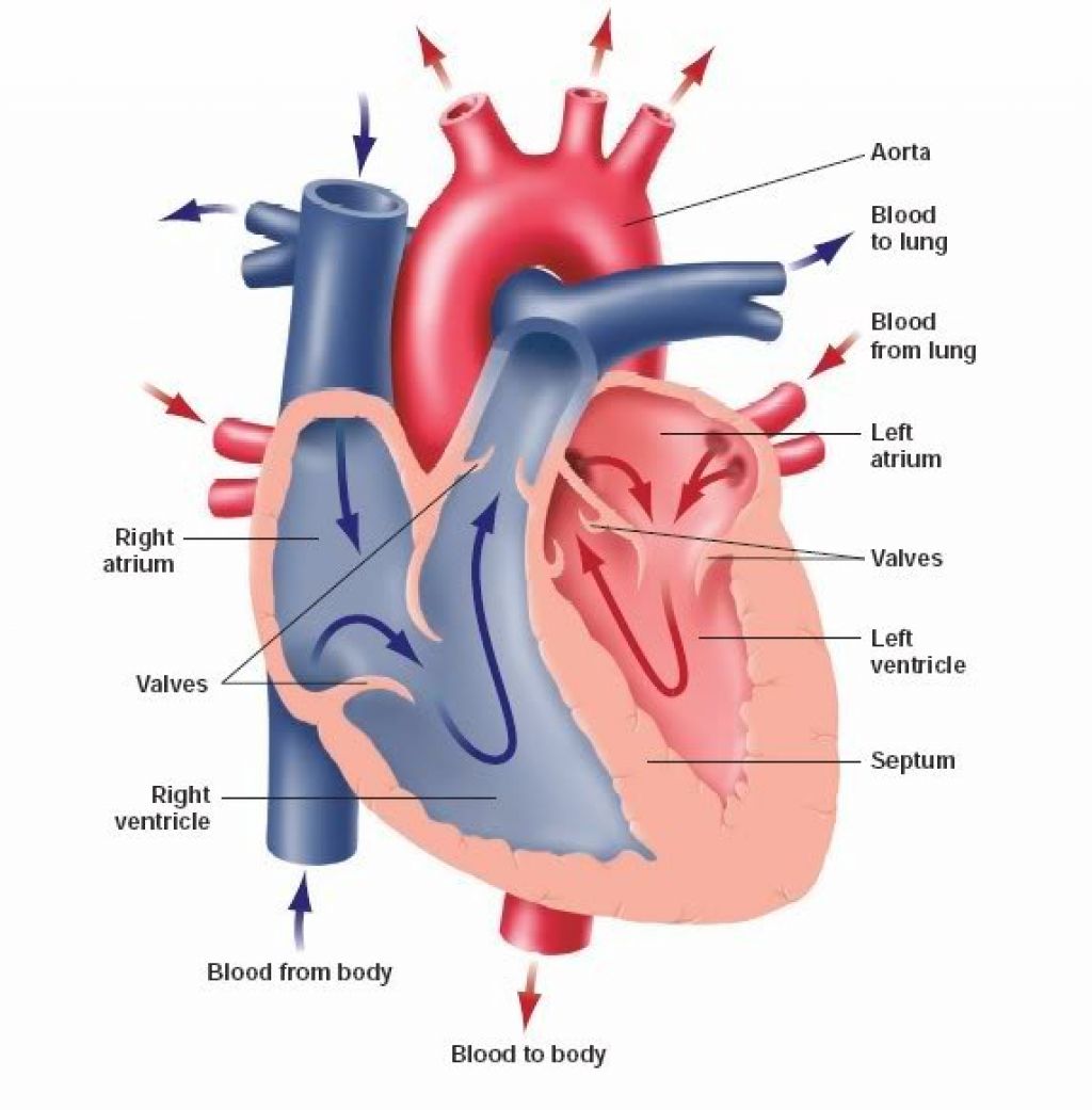
The heart The heart is a unidirectional pump. Valves are present to prevent the backflow of blood. The right side pumps deoxygenated blood (low in oxygen and high in carbon dioxide) to the.
Simple Human Heart Diagram Clipart Free Clip Art Images Cliparts.co
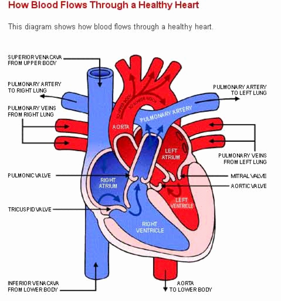
Blood flow through the heart made easy! This video provides a simple step-by-step diagram of the cardiac blood flow and a chart of the circulation pathway. Includes the anatomy of the heart and an animation quiz at the end in order to test your knowledge! Save Time with a Video!
Human Heart Simple Drawing at GetDrawings Free download
Selecting or hovering over a box will highlight each area in the diagram. For optimal viewing of this interactive, view at your screen's default zoom setting (100%) and with your browser window view maximised. See the Labelling the heart activity for additional support in using this interactive. Parts of the heart
Heart Blood Flow Simple Anatomy Diagram, Cardiac Circulation Pathway Steps — EZmed
Welcome to the anatomy of the heart made easy! We will use labeled diagrams and pictures to learn the main cardiac structures and related vascular system. In addition to reviewing the human heart anatomy, we will also discuss the function and order in which blood flows through the heart.
When one teaches, two learn. The heart and the circulatory system (DIAGRAMS)
Myocardium - a thick, muscular middle layer that contracts and relaxes to pump blood around of your heart. Endocardium - a thin, inner layer that makes up the lining of the four chambers and the valves in your heart. What does the heart's electrical system do?